

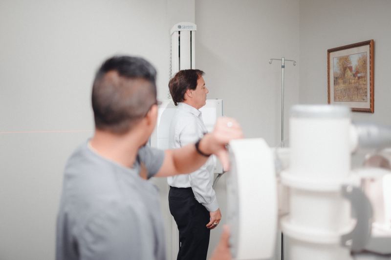
Fluoroscopy for Car Accident Injuries in Oklahoma City
Fluoroscopy is X-ray in motion. Our two GE 9800 C-arm systems let your physician watch the spine and joints move in real time and place injection needles precisely where they need to go. Fluoroscopy is scheduled as its own appointment at Accident Care.
- Real-time X-ray video
- Image-guided injections
- Pulsed, low-dose modes
- 2GE 9800 C-arm systems
- LiveMoving X-ray image
- PulsedDose-saving fluoroscopy mode
- ScheduledSeparate appointment
Why fluoroscopy matters after a crash
What is fluoroscopy?
Fluoroscopy is a real-time X-ray technique that shows a continuous moving image on a monitor. A C-arm fluoroscope holds the X-ray source and image receptor on opposite ends of a C-shaped arm that rotates around the patient. After a car accident, fluoroscopy is used to guide spinal and joint injections with needle-tip accuracy and to watch how the spine and joints move.
Some injuries are best understood in motion, and some treatments need to be delivered to an exact spot millimeters from a nerve. Fluoroscopy serves both. It shows bones and needle position live, so a physician can confirm a needle is in the correct joint or epidural space before any medication is injected.
Accident Care has two GE 9800 C-arm fluoroscopy systems on-site. Fluoroscopic procedures are scheduled as a separate appointment after your evaluation, so your physician can plan the procedure and review your imaging and medical history first.
How fluoroscopy works
An X-ray tube on one end of the C-arm sends a low-intensity beam through the body to an image intensifier on the other end. The intensifier converts the X-ray pattern into a bright visible image, which a camera displays on the monitor in real time.
Because the source and receptor are fixed to the same arm, the C-arm can rotate around you to give AP (front), lateral (side), and oblique views without moving you. Oblique views are how physicians line up the facet joints or the neural foramina for needle placement.
Pulsed fluoroscopy fires short bursts of X-rays instead of a continuous beam, which reduces dose while keeping the image usable. “Last image hold” keeps the final frame on screen so the physician can study it without more exposure, and tight collimation limits the beam to the area of interest.
For injections, a small amount of iodinated contrast is often given through the needle before the medication. Its spread pattern confirms the needle tip is in the intended space and helps detect unintended flow into a blood vessel, a key safety step.
How fluoroscopy is used for spine and joint care
| Use | What fluoroscopy adds |
|---|---|
| Epidural steroid injections (interlaminar or transforaminal) | Confirms the needle path and shows contrast spreading in the epidural space along the targeted nerve root |
| Facet joint injections and medial branch blocks | Lines up the target joint or nerve on oblique views; diagnostic blocks help confirm whether a facet joint is the source of pain |
| Sacroiliac joint injections | Shows the narrow joint line and confirms intra-articular contrast |
| Large-joint injections | Guides the needle into the shoulder, hip, or knee joint when landmarks are difficult |
| Dynamic spine and joint imaging | Watches vertebrae or a joint move through a range of motion to look for abnormal motion |
What fluoroscopy helps your physician do
Precise needle placement
Millimeter-level guidance near nerves and joints.
Image-guided spinal injections are the accepted standard of care because landmarks alone are not reliable for reaching the epidural space or facet joints accurately.
Identify the pain generator
Diagnostic blocks confirm the source of pain.
If numbing a specific facet joint or nerve briefly relieves your pain, that points to it as the cause and guides further treatment.
Spine in motion
Real-time view of segmental motion.
Watching vertebrae move can reveal abnormal motion between levels that a single still image cannot show.
Safer injections
Contrast confirms where medication will go.
Contrast checks before injecting medication help avoid injection into a blood vessel and confirm the medication will reach the inflamed area.
Hurt in a car accident? See a medical doctor. No outside imaging center, no upfront out-of-pocket cost.
What Fluoroscopy can and can’t do
Strengths
- Real-time imaging: the physician sees the needle and anatomy as they move
- C-arm rotation gives multiple angles without repositioning you
- Pulsed modes and last-image-hold keep dose low
- Contrast confirmation adds a safety check before medication is given
Limits
- Uses ionizing radiation, so it is used only when it adds value
- Shows bone and contrast, not soft tissue detail; MRI is used for that
- Contrast is iodine-based, so allergies must be reviewed first
- Requires a scheduled appointment rather than a walk-in study
Before a fluoroscopy-guided procedure
- Tell us if you are or may be pregnant.
- Tell us about any prior reaction to iodinated contrast. (A shellfish allergy on its own is not a reason to avoid contrast, but mention any allergies.)
- Tell us about blood thinners (anticoagulants or antiplatelet drugs). Some must be managed before spinal injections, and only with guidance from the prescribing physician.
- Tell us about diabetes; steroid injections can temporarily raise blood sugar.
- Staff wear lead protection and use collimation and pulsed modes to keep everyone’s exposure as low as reasonably achievable.
Step by step at Accident Care
- 1
Evaluation and planning
Your physician reviews your exam and any MRI or CT to decide whether a guided procedure or dynamic study is appropriate.
- 2
Scheduled appointment
Fluoroscopy is scheduled as its own appointment here at Accident Care.
- 3
Preparation
You review medications and allergies, and the skin over the target area is cleaned and numbed.
- 4
Guided procedure
The C-arm is positioned for the views needed, and the physician advances the needle under live X-ray guidance, confirming position with contrast before giving medication.
- 5
Recovery
You are monitored briefly afterward and given activity and pain-tracking instructions.
- 6
Follow-up
Your doctor reviews your response at your next appointment and adjusts your treatment plan.
Fluoroscopy after a car accident: common questions
What is the difference between fluoroscopy and a regular X-ray?
A regular X-ray is a single still image. Fluoroscopy is a continuous, real-time X-ray image, like a video, used to watch motion or guide a needle while the procedure is happening.
Why are spinal injections done with fluoroscopy?
The epidural space, facet joints, and nerve roots are small and deep. Fluoroscopy shows the needle tip relative to the bones in real time, and contrast confirms where the medication will flow, which improves accuracy and safety compared with landmark-only injections.
How much radiation is involved?
Fluoroscopy time for a typical spinal injection is short, and pulsed modes and last-image-hold reduce dose further. Your physician uses the least exposure needed to place the needle safely.
Can I get fluoroscopy the same day as my first visit?
No. Fluoroscopy is scheduled as a separate appointment so your physician can review your imaging, medications, and history and plan the procedure.
I’m allergic to contrast. Can I still have a guided injection?
Tell your physician. Depending on the reaction you had, options include premedication, alternative approaches, or performing the procedure without contrast in some cases. Your physician will decide what is safe for you.
Does insurance cover fluoroscopy-guided injections after a car accident?
When another driver caused the accident, their insurance covers medically necessary treatment, including image-guided procedures. Accident Care bills the insurance directly, so there is zero upfront out-of-pocket cost to you.
Does a fluoroscopy-guided injection hurt?
The skin is numbed with local anesthetic first. Most people feel pressure and brief discomfort as the needle is positioned, and sometimes a temporary reproduction of their usual pain when the medication reaches the target.
How long does a fluoroscopy-guided spinal injection take?
The procedure itself often takes 15 to 30 minutes. Plan extra time for preparation beforehand and a short monitoring period afterward.
How soon does an epidural steroid injection start working?
The local anesthetic can bring relief within minutes but wears off within hours. The steroid usually takes a few days to start reducing inflammation, and its full effect may take up to about two weeks.
What are the side effects of a spinal injection?
Common, temporary effects include soreness at the injection site, facial flushing, trouble sleeping, and raised blood sugar in people with diabetes. Serious complications such as infection, bleeding, or nerve injury are rare. Your physician reviews the risks with you before the procedure.
What is a medial branch block?
A medial branch block numbs the small nerves that carry pain signals from a facet joint. If your pain drops significantly afterward, that points to the facet joint as the pain generator and guides further treatment.
How many steroid injections can I have?
There is no single limit for everyone. Physicians space injections and limit total steroid exposure based on your response, your health, and the medication used. Your physician decides with you whether repeating an injection makes sense.
Should I stop my blood thinner before an injection?
Never stop a blood thinner on your own. Some must be paused before certain spinal injections, but only with guidance from the physician who prescribes it. Tell your Accident Care physician about every medication you take.
Other diagnostic tools at Accident Care
MRI
How our on-site GE 1.5 Tesla wide-bore MRI finds disc herniations, ligament tears, bone bruises and nerve compression after a crash.
Learn moreCT scan
How our on-site Siemens SOMATOM go.Top 128-slice CT finds fractures and bony injuries after a crash.
Learn moreUltrasound
How our on-site Canon Aplio a550 ultrasound finds muscle, tendon, and ligament injuries after a crash, in real time and without radiation.
Learn moreX-ray
How digital X-ray is used after a crash: cervical and lumbar series, flexion-extension views, alignment lines, instability criteria, radiation dose, and what X-rays can and can’t show.
Learn moreNerve testing (EMG/NCV)
How EMG and nerve conduction velocity (NCV) studies diagnose pinched nerves, radiculopathy, and nerve injury after a crash: latency, amplitude, conduction velocity, denervation timing, and what to expect.
Learn moreFunctional & lab tests
How Accident Care measures injury objectively: range of motion in degrees, strength grading, reflexes, provocative tests, validated disability scores like the NDI and Oswestry, and targeted lab work.
Learn more
Sources and further reading
- Spine Intervention Society. Practice Guidelines for Spinal Diagnostic and Treatment Procedures, 2nd ed.
- Rathmell JP, et al. Safeguards to prevent neurologic complications after epidural steroid injections: consensus opinions from a multidisciplinary working group. Anesthesiology, 2015.
- U.S. Food and Drug Administration. Fluoroscopy: radiation-emitting products and procedures.
- American College of Radiology. ACR Manual on Contrast Media.
This page is educational and doesn’t replace an evaluation by a physician. Last reviewed November 2026. If you have severe headache, confusion, weakness, loss of bladder or bowel control, or chest or abdominal pain after a crash, call 911 or go to the nearest emergency room.
Get the answers you need, all in one place.
Same-day appointments available. Walk-ins welcome. No upfront out-of-pocket cost.