

Musculoskeletal Ultrasound After a Car Accident in Oklahoma City
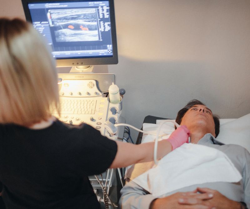
Our Canon Aplio a550 ultrasound system lets your physician watch muscles, tendons, and ligaments move in real time, with no radiation. It is ideal for shoulder, knee, ankle, wrist, and muscle injuries from a crash, and it is on-site at Accident Care.
- Real-time, dynamic imaging
- No radiation
- Side-to-side comparison
- Real timeWatch tissue move during the exam
- High MHzLinear probes for fine tendon detail
- 15–30 minTypical study
- 0 mSvIonizing radiation
Why ultrasound matters after a crash
What is diagnostic ultrasound?
Diagnostic ultrasound sends high-frequency sound waves into the body and builds an image from the echoes that return. Musculoskeletal (MSK) ultrasound uses high-frequency linear transducers to show tendons, ligaments, muscles, bursae, and superficial nerves in fine detail and in motion. After a car accident it is used to find tendon and ligament tears, muscle strains and hematomas, bursitis, and joint effusions, without radiation.
Bracing against the steering wheel, a seatbelt across the shoulder, or a knee against the dashboard can tear or strain the soft tissues around joints. Ultrasound looks at those structures directly, and unlike any other imaging test it can show them moving: a tendon gliding, a ligament under stress, or a tendon snapping out of its groove.
Accident Care uses a Canon Aplio a550 system. Your physician orders ultrasound when your exam points to a specific soft-tissue injury near the surface, and the findings are read and reviewed with you at your next appointment, usually within a week.
How ultrasound works
The transducer contains piezoelectric crystals that turn electrical pulses into sound waves far above human hearing, and turn the returning echoes back into electrical signals. The system times each echo to calculate depth and builds a gray-scale image many times per second.
Echoes form wherever sound crosses a boundary between tissues with different acoustic impedance. Radiologists describe what they see as hyperechoic (bright, such as a normal tendon or bone surface), hypoechoic (darker, such as fluid-filled or swollen tissue), or anechoic (black, such as a fluid collection).
Frequency is a trade-off. Higher frequencies give finer resolution but do not travel as deep. MSK studies use high-frequency linear transducers for superficial tendons and ligaments, and lower-frequency curved probes for deeper structures such as the hip or large muscles.
Doppler imaging detects motion of red blood cells. Color and power Doppler show increased blood flow (hyperemia) in inflamed tendons and healing tissue. Canon’s Superb Micro-vascular Imaging (SMI) on the Aplio platform is designed to show very slow, small-vessel flow that conventional Doppler can miss.
A well-known MSK artifact, anisotropy, makes a normal tendon look falsely dark if the beam is not perpendicular to its fibers. Experienced examiners rock the probe to rule it out, one of the reasons ultrasound is called operator-dependent.
Common post-accident ultrasound studies
| Area | What is evaluated |
|---|---|
| Shoulder | Rotator cuff (supraspinatus, infraspinatus, subscapularis), biceps tendon, subacromial bursa, AC joint, dynamic impingement |
| Knee | Quadriceps and patellar tendons, collateral ligaments, joint effusion, Baker’s cyst |
| Ankle and foot | Anterior talofibular and calcaneofibular ligaments, Achilles and peroneal tendons, tendon subluxation |
| Wrist and elbow | Tendons, ganglion cysts, median nerve at the carpal tunnel, ulnar nerve at the elbow |
| Muscle | Strain grading, tears, intramuscular hematoma, and its evolution over time |
What ultrasound finds after a car accident
Rotator cuff tears
Partial- and full-thickness tears.
In skilled hands, ultrasound performs comparably to MRI for full-thickness rotator cuff tears, and it can compare the injured shoulder with the other side in the same visit.
Muscle strain and hematoma
Tears, fluid collections, and bleeding within muscle.
Seatbelt and impact injuries can cause intramuscular bleeding. Ultrasound shows its size and can be repeated to confirm it is resolving.
Tendon injury
Tendinopathy, partial tears, ruptures, and subluxation.
Dynamic scanning can catch a peroneal or biceps tendon slipping out of its groove, something static imaging can miss.
Ligament sprains
Ankle, knee, and elbow ligaments.
Ultrasound shows thickening, fiber disruption, and laxity when the joint is gently stressed during the exam.
Bursitis and effusion
Fluid in bursae and joints.
Post-traumatic bursitis and joint effusions are easy to see and measure.
Peripheral nerves
Swelling and entrapment of superficial nerves.
The median nerve’s cross-sectional area at the carpal tunnel and the ulnar nerve at the elbow can be measured, complementing nerve conduction studies.
Hurt in a car accident? See a medical doctor. No outside imaging center, no upfront out-of-pocket cost.
What Ultrasound can and can’t do
Strengths
- No ionizing radiation; safe to repeat and safe in pregnancy
- Dynamic: shows tissue moving and under stress
- Immediate side-to-side comparison with your uninjured side
- Comfortable and works around most metal implants
Limits
- Sound cannot pass through bone, so it can’t see inside joints or the spinal canal
- Not used to evaluate spinal discs or nerve roots; MRI does that
- Deep structures are harder to see in larger body types
- Operator-dependent: image quality depends on the examiner’s technique
Ultrasound safety
- Diagnostic ultrasound uses no ionizing radiation.
- It is routinely used in pregnancy and can be repeated as often as needed.
- Water-based gel helps the sound waves pass from the probe into your skin. It wipes off afterward.
- Gentle pressure from the probe may be uncomfortable over an injured area; tell the examiner.
Step by step at Accident Care
- 1
Your physician orders the study
Ultrasound is ordered when your exam points to a specific tendon, ligament, muscle, or bursa injury.
- 2
Preparation
No special preparation. Wear clothing that lets us reach the area being examined.
- 3
The exam
Gel is applied and the transducer is moved over the area. You may be asked to move the joint so the examiner can watch the tissue in motion.
- 4
Comparison and Doppler
The uninjured side is often scanned for comparison, and Doppler is used to look at blood flow.
- 5
Interpretation
Images and measurements are interpreted and a written report is issued.
- 6
Results with your doctor
Your doctor reviews the results with you at your next appointment, usually within a week.
Ultrasound after a car accident: common questions
Is ultrasound as good as an MRI?
For some questions, yes. Ultrasound is comparable to MRI for full-thickness rotator cuff tears and many superficial tendon and ligament injuries, and it adds real-time motion. MRI is better for deep structures, the inside of joints (such as the labrum, meniscus, and ACL), the spine, and bone marrow.
Can ultrasound show a herniated disc?
No. Sound waves cannot pass through the bones of the spine. Disc herniations and nerve root compression are evaluated with MRI.
Does an ultrasound hurt?
No. You may feel gentle pressure from the probe, which can be tender over an injured area. There are no needles and no radiation.
Why does the examiner ask me to move during the scan?
Dynamic scanning is one of ultrasound’s biggest advantages. Moving the joint shows how a tendon glides, whether it slips out of place, or whether a ligament opens under gentle stress, findings that a still image can miss.
What is Doppler ultrasound?
Doppler detects blood flow. In injured tendons and soft tissue, increased flow can indicate active inflammation or healing, and it helps tell a fluid collection apart from solid tissue.
Does insurance cover an ultrasound after a car accident?
When another driver caused the accident, their insurance covers medically necessary imaging, including ultrasound. Accident Care bills the insurance directly, so there is zero upfront out-of-pocket cost to you.
How long does a musculoskeletal ultrasound take?
Most studies take 15 to 30 minutes, depending on how many structures are examined and whether the other side is scanned for comparison.
How do I prepare for an ultrasound?
No special preparation is needed. Wear loose clothing that lets us reach the area, such as a tank top for a shoulder study or shorts for a knee study.
Can ultrasound show a torn ACL or meniscus?
Only partly. Ultrasound sees structures near the surface well, but the ACL and meniscus sit deep inside the knee joint. MRI is the test for those injuries.
Can ultrasound show a bruise or blood collection in a muscle?
Yes. Ultrasound shows intramuscular hematomas clearly, measures their size, and can be repeated to confirm they are resolving.
Is ultrasound safe during pregnancy?
Yes. Diagnostic ultrasound uses sound waves, not ionizing radiation, and is routinely used in pregnancy.
Is ultrasound better than MRI for a shoulder injury?
For full-thickness rotator cuff tears, ultrasound performs comparably to MRI in skilled hands and adds real-time motion. MRI is better for the labrum, cartilage, and bone marrow, so your physician chooses based on the suspected injury.
Can ultrasound see nerves?
Yes, superficial nerves such as the median nerve at the wrist and the ulnar nerve at the elbow. Swelling of the median nerve at the carpal tunnel can be measured, which complements nerve conduction testing.
Why does the examiner scan my uninjured side too?
Comparing the injured side with your normal side shows what is normal for your body, making subtle thickening, tears, or fluid easier to identify.
When will I get my ultrasound results?
The study is interpreted and a written report is issued, and your doctor reviews the results with you at your next appointment, usually within a week.
Other diagnostic tools at Accident Care
MRI
How our on-site GE 1.5 Tesla wide-bore MRI finds disc herniations, ligament tears, bone bruises and nerve compression after a crash.
Learn moreCT scan
How our on-site Siemens SOMATOM go.Top 128-slice CT finds fractures and bony injuries after a crash.
Learn moreX-ray
How digital X-ray is used after a crash: cervical and lumbar series, flexion-extension views, alignment lines, instability criteria, radiation dose, and what X-rays can and can’t show.
Learn moreFluoroscopy
How Accident Care uses two GE 9800 C-arm fluoroscopy systems for real-time spine and joint imaging and image-guided injections.
Learn moreNerve testing (EMG/NCV)
How EMG and nerve conduction velocity (NCV) studies diagnose pinched nerves, radiculopathy, and nerve injury after a crash: latency, amplitude, conduction velocity, denervation timing, and what to expect.
Learn moreFunctional & lab tests
How Accident Care measures injury objectively: range of motion in degrees, strength grading, reflexes, provocative tests, validated disability scores like the NDI and Oswestry, and targeted lab work.
Learn more
Sources and further reading
- American Institute of Ultrasound in Medicine (AIUM). Practice Parameter for the Performance of a Musculoskeletal Ultrasound Examination.
- Roy JS, et al. Diagnostic accuracy of ultrasonography, MRI and MR arthrography in the characterisation of rotator cuff disorders: a systematic review and meta-analysis. British Journal of Sports Medicine, 2015.
- American College of Radiology. ACR–AIUM–SPR–SRU Practice Parameter for the Performance of the Musculoskeletal Ultrasound Examination.
This page is educational and doesn’t replace an evaluation by a physician. Last reviewed November 2026. If you have severe headache, confusion, weakness, loss of bladder or bowel control, or chest or abdominal pain after a crash, call 911 or go to the nearest emergency room.
Get the answers you need, all in one place.
Same-day appointments available. Walk-ins welcome. No upfront out-of-pocket cost.