

MRI After a Car Accident: 1.5 Tesla Wide-Bore Imaging in Oklahoma City
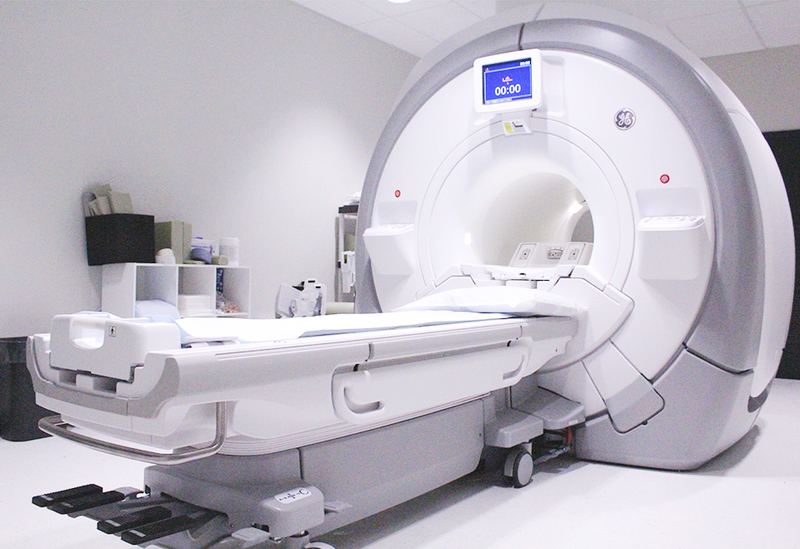
Our GE Signa 450W 1.5 Tesla wide-bore MRI shows the injuries X-rays can’t: disc herniations, ligament tears, bone bruises, and nerve compression. It is on-site at 3209 NW Expressway and ordered by your Accident Care physician, so there is no outside imaging center to coordinate with.
- 1.5 Tesla high-field
- Wide, short bore
- No ionizing radiation
- 1.5 TMagnetic field strength
- 63.9 MHzHydrogen resonance frequency at 1.5 T
- 30–45 minTypical cervical or lumbar study
- 0 mSvIonizing radiation
Why MRI matters after a crash
What is an MRI?
An MRI (magnetic resonance imaging) scan uses a strong magnetic field and radiofrequency pulses, not X-rays, to map the hydrogen protons in water and fat. The result is cross-sectional images with far better soft-tissue contrast than X-ray or CT. After a car accident, MRI is the reference standard for diagnosing disc herniation, ligament injury, spinal cord and nerve root compression, and bone marrow edema (bone bruising).
Most car accident injuries are soft-tissue injuries: discs, ligaments, muscles, tendons, joint capsules, and nerves. X-ray and CT are built to show bone. MRI is the only routine imaging test that shows those soft tissues in fine detail and can also show bone marrow edema, the “bone bruise” pattern that marks recent trauma.
At Accident Care, MRI is ordered by the physician who examined you, based on your exam findings, not as a blanket screen. The scan is performed in our own building on a GE Signa 450W, a 1.5 Tesla high-field magnet with a wide, short bore. MRI is scheduled as its own appointment, board-certified radiologists read every study, and your doctor goes over the results with you at your next appointment, usually within a week.
How MRI works, step by step
Hydrogen nuclei (single protons) behave like tiny bar magnets. Inside the 1.5 tesla field, a little more than half of them line up with the field, and all of them wobble (precess) at the Larmor frequency: 42.58 MHz per tesla, or about 63.9 MHz at 1.5 T.
A radiofrequency pulse tuned to exactly that frequency tips the protons out of alignment. When the pulse stops, they relax back and give off a faint radio signal that is picked up by receiver coils placed over the body part being scanned.
Tissues relax at different rates. T1 relaxation (how fast protons realign with the field) and T2 relaxation (how fast their signal dephases) differ between fat, water, disc, ligament, cord, and marrow. By changing the pulse timing (repetition time, TR, and echo time, TE), the scanner weights the image toward one property or the other. That is where image contrast comes from.
Gradient coils briefly vary the field across the body so each signal can be traced back to its exact location. Switching those gradients on and off is what makes the loud knocking sound during a scan.
Signal-to-noise ratio rises roughly with field strength. 1.5 T is the long-standing clinical standard for spine and musculoskeletal imaging. Low-field “open” scanners, often 0.2 to 0.3 T, trade image quality for openness, which makes subtle annular fissures, ligament injuries, and early marrow edema easier to miss. A wide-bore 1.5 T design keeps high-field image quality while giving you more room.
MRI sequences and what each one shows
| Sequence | What it shows after an accident |
|---|---|
| T1-weighted | Anatomy and marrow. Fat is bright. Shows fracture lines, marrow replacement, and chronic changes such as fatty endplate (Modic type 2) change. |
| T2-weighted | Fluid is bright. Shows disc hydration, herniations, spinal canal and foraminal narrowing, cerebrospinal fluid, and spinal cord signal. |
| STIR (short tau inversion recovery) | Suppresses fat so edema stands out. The key sequence for bone bruises, acute compression fractures, ligament sprains, and muscle strain. |
| Fat-saturated T2 / proton density | Joint imaging: rotator cuff, labrum, meniscus, and cruciate and collateral ligament tears. |
| Gradient echo / susceptibility-weighted (SWI) | Very sensitive to blood products, including the microhemorrhages of diffuse axonal injury in traumatic brain injury. |
| Diffusion-weighted (DWI) | Measures water motion. Used in brain imaging and to help characterize some marrow and soft-tissue lesions. |
What MRI finds after a car accident
Disc injury
Protrusion, extrusion, sequestration, and annular fissures.
Radiologists use the standard NASS/ASNR lumbar disc nomenclature. A protrusion is a broad-based displacement; an extrusion has a neck narrower than its dome; a sequestered fragment has lost continuity with the parent disc. A bright spot in the annulus on T2 (a “high-intensity zone”) marks an annular fissure.
Bone marrow edema
Bone bruises and occult fractures, best seen on STIR.
Edema in a vertebral body, endplate, or joint surface points to recent impact. It is often invisible on X-ray and CT, which is why it matters for diagnosis and documentation.
Ligament and facet injury
Spinal and joint ligaments, facet capsules, and effusions.
Includes the anterior and posterior longitudinal ligaments, interspinous ligaments, the alar and transverse ligaments at the top of the neck, and facet joint effusions after flexion-extension (whiplash) loading.
Nerve and cord compression
Foraminal and central canal stenosis, cord signal change.
MRI shows exactly where a disc or osteophyte contacts a nerve root, and whether the spinal cord itself shows abnormal T2 signal (myelopathy), which changes how urgently care is needed.
Joint and soft-tissue tears
Shoulder, knee, hip, and wrist injuries.
Rotator cuff and labral tears from bracing on the steering wheel, meniscus and ACL injuries from dashboard impact, and muscle strains or hematomas.
Traumatic brain injury
Contusions and diffuse axonal injury.
Susceptibility-weighted imaging can detect microbleeds that CT misses. A normal MRI does not rule out a concussion, which is a clinical diagnosis, but MRI helps when symptoms persist or the exam raises concern.
How MRI helps separate acute injury from old degeneration
Insurers often argue that findings on an MRI were “already there.” MRI can help answer that question because acute injury and chronic degeneration look different.
Signs that point to recent injury include bone marrow edema on STIR, prevertebral or paraspinal soft-tissue edema, ligament edema, and new joint effusions. Signs of long-standing degeneration include disc desiccation (dark discs on T2), osteophytes, endplate sclerosis, and Modic type 2 or 3 endplate changes. Modic type 1 change is itself an edema pattern and is interpreted in context.
Degenerative findings are common in people with no pain at all. In a large systematic review (Brinjikji et al., AJNR 2015), disc bulges were found in about 30% of pain-free 20-year-olds and 84% of pain-free 80-year-olds. That is why your physician never reads an MRI in isolation: the report is correlated with your exam, your symptoms, and the timeline of your accident. Many crash patients have both, and documenting both lets your physician explain whether the crash caused a new injury or aggravated an existing condition.
Hurt in a car accident? See a medical doctor. No outside imaging center, no upfront out-of-pocket cost.
What MRI can and can’t do
Strengths
- Best soft-tissue contrast of any imaging test: discs, ligaments, cord, nerves, muscle, cartilage
- Shows bone marrow edema and occult fractures that X-ray and CT miss
- No ionizing radiation, so it can be repeated to track healing
- Images in any plane (sagittal, axial, coronal) without moving you
Limits
- Longer than X-ray or CT; motion blurs the images, so you need to hold still
- Some implants and metal fragments are unsafe or distort the images
- Fine cortical bone detail and complex fractures are better seen on CT
- Age-related findings are common in people without pain and must be correlated with your exam
MRI safety screening
- Everyone completes a safety questionnaire before entering the scanner room. The magnet is always on.
- Tell us about pacemakers, defibrillators, neurostimulators, cochlear implants, insulin pumps, aneurysm clips, or any implanted device. Many modern devices are “MR-conditional” and can be scanned under specific conditions; bring your device card.
- If you have ever had metal in your eyes (welding, grinding), a screening X-ray of the orbits may be needed first.
- Most post-accident spine and joint MRIs are non-contrast. If gadolinium contrast is ever needed, kidney function and allergy history are reviewed first.
- Tell us if you are or may be pregnant. Non-contrast MRI is generally considered acceptable in pregnancy when clinically needed, but your physician makes that decision with you.
Step by step at Accident Care
- 1
Your physician orders the study
After your exam, your Accident Care physician decides whether MRI is medically necessary and which body region and sequences to order.
- 2
Your MRI appointment is scheduled
MRI is scheduled as its own appointment here in our facility. No outside imaging center and no separate referral.
- 3
Safety screening and preparation
You complete the safety questionnaire, remove metal objects, and may change into a gown. Tell the technologist if you are anxious about enclosed spaces.
- 4
The scan
You lie on the table with a receiver coil over the area being imaged. You get hearing protection for the knocking sounds. A typical cervical or lumbar study takes 30 to 45 minutes.
- 5
Radiologist interpretation
A board-certified radiologist reviews every sequence and issues a written report.
- 6
Results with your doctor
Your doctor reviews the images and report with you at your next appointment, usually within a week, and updates your treatment plan.
MRI after a car accident: common questions
What does an MRI show that an X-ray does not?
X-ray shows bone and alignment. MRI shows the soft tissues injured in most car accidents: discs, ligaments, muscles, tendons, cartilage, the spinal cord, and nerve roots. It also shows bone marrow edema (bone bruising) and some fractures that are invisible on X-ray.
Is a 1.5 Tesla MRI good enough, or do I need 3 Tesla?
1.5 T is the established clinical standard for spine and musculoskeletal imaging. 3 T offers more signal, which helps in some brain and small-joint studies, but it also produces more artifact near metal hardware and more tissue heating. For most post-accident spine and joint questions, a high-field 1.5 T scanner answers the clinical question.
Will an MRI show whiplash?
Whiplash (whiplash-associated disorder) is a clinical diagnosis based on the mechanism of injury and your exam. In milder grades the MRI is often normal. MRI is used to look for the injuries that can come with whiplash: disc herniation, ligament injury, facet effusion, bone bruising, and nerve root compression.
How soon after a car accident should I get an MRI?
Not everyone needs one right away. Your physician orders MRI when the exam suggests a disc, ligament, or nerve injury, when there are neurological signs such as numbness or weakness, or when symptoms are not improving as expected. Red-flag symptoms are imaged sooner.
I’m claustrophobic. Can I still have an MRI?
Usually, yes. Our GE Signa 450W has a wider, shorter bore than older scanners, so there is more room around your head and body. Tell the team ahead of time; we can talk you through the scan, and in some cases your physician can discuss options to help you relax.
Do I need contrast dye for my MRI?
Most MRIs after a car accident do not need contrast. Gadolinium contrast is reserved for specific questions, such as a suspected infection, tumor, or certain post-surgical evaluations. Your physician will tell you if it is needed.
Can an MRI tell whether my injury is new?
Often it can help. Bone marrow edema, soft-tissue edema, and ligament edema point to a recent injury, while disc desiccation, osteophytes, and certain endplate changes point to long-standing degeneration. Your physician correlates those findings with your exam and the date of your accident.
Does insurance cover an MRI after a car accident in Oklahoma?
When another driver caused the accident, their insurance covers medically necessary imaging, including MRI. Accident Care bills the insurance directly, so there is zero upfront out-of-pocket cost to you.
How long does an MRI take?
A typical cervical or lumbar MRI takes 30 to 45 minutes. Scanning more than one region, such as the neck and lower back, takes longer. Plan a little extra time for safety screening and changing.
Why is the MRI so loud?
The knocking and buzzing come from the gradient coils switching on and off rapidly to encode where each signal comes from. It is normal. You will get earplugs or headphones.
Can I have an MRI if I have metal plates, screws, or a joint replacement?
Usually, yes. Most modern orthopedic implants are MR-safe or MR-conditional. They can distort the image right next to the metal, so tell us about every implant and surgery during screening.
Can I have an MRI if I have tattoos?
Generally, yes. Rarely, some tattoo inks or permanent makeup cause warmth or tingling during the scan. Tell the technologist right away if you feel heat at a tattoo site.
Do I need to do anything to prepare for my MRI?
For a non-contrast MRI, you can usually eat, drink, and take your medications as normal. Leave jewelry at home, avoid clothing with metal, and complete the safety questionnaire honestly.
What is the difference between a disc bulge and a disc herniation?
Under the standard NASS/ASNR nomenclature, a bulge is a generalized extension of the disc beyond its normal margins involving more than 25% of its circumference. A herniation is localized, involving less than 25%, and is further described as a protrusion, extrusion, or sequestration.
What does an “annular fissure” or “annular tear” on my MRI mean?
It is a separation in the fibers of the disc’s tough outer ring, often seen as a bright spot on T2 images. Fissures can be degenerative or related to injury, so your physician interprets the finding alongside your symptoms and the timing of your accident.
My MRI report says “no acute findings,” but I’m still in pain. Why?
Many real injuries, such as muscle strains, facet joint sprains, and some ligament injuries, are not visible on MRI. A normal or non-acute MRI doesn’t mean you aren’t hurt. Your diagnosis is based on your exam and history as well as imaging.
Will I get my MRI results right away?
No. A board-certified radiologist reviews every sequence and issues a written report, and your doctor reviews the results with you at your next appointment, usually within a week.
Can an MRI detect a concussion or traumatic brain injury?
A standard MRI is often normal after a concussion, which is a clinical diagnosis. Sequences such as susceptibility-weighted imaging can detect microbleeds from more significant traumatic brain injuries, so MRI is ordered when symptoms persist or the exam raises concern.
Do I need a referral to get an MRI at Accident Care?
No outside referral is needed. Your Accident Care physician orders the MRI after examining you, and it is performed in our own building.
Other diagnostic tools at Accident Care
CT scan
How our on-site Siemens SOMATOM go.Top 128-slice CT finds fractures and bony injuries after a crash.
Learn moreUltrasound
How our on-site Canon Aplio a550 ultrasound finds muscle, tendon, and ligament injuries after a crash, in real time and without radiation.
Learn moreX-ray
How digital X-ray is used after a crash: cervical and lumbar series, flexion-extension views, alignment lines, instability criteria, radiation dose, and what X-rays can and can’t show.
Learn moreFluoroscopy
How Accident Care uses two GE 9800 C-arm fluoroscopy systems for real-time spine and joint imaging and image-guided injections.
Learn moreNerve testing (EMG/NCV)
How EMG and nerve conduction velocity (NCV) studies diagnose pinched nerves, radiculopathy, and nerve injury after a crash: latency, amplitude, conduction velocity, denervation timing, and what to expect.
Learn moreFunctional & lab tests
How Accident Care measures injury objectively: range of motion in degrees, strength grading, reflexes, provocative tests, validated disability scores like the NDI and Oswestry, and targeted lab work.
Learn more
Sources and further reading
- Fardon DF, et al. Lumbar disc nomenclature: version 2.0. Recommendations of the combined task forces of NASS, ASSR, and ASNR. The Spine Journal, 2014.
- Brinjikji W, et al. Systematic literature review of imaging features of spinal degeneration in asymptomatic populations. AJNR American Journal of Neuroradiology, 2015.
- American College of Radiology. ACR Manual on MR Safety.
- American College of Radiology. ACR Appropriateness Criteria: Low Back Pain; Cervical Neck Pain or Cervical Radiculopathy.
- Spitzer WO, et al. Scientific monograph of the Quebec Task Force on Whiplash-Associated Disorders. Spine, 1995.
This page is educational and doesn’t replace an evaluation by a physician. Last reviewed November 2026. If you have severe headache, confusion, weakness, loss of bladder or bowel control, or chest or abdominal pain after a crash, call 911 or go to the nearest emergency room.
Get the answers you need, all in one place.
Same-day appointments available. Walk-ins welcome. No upfront out-of-pocket cost.